Let Us Know About Fracture of the Condyles of the Tibia

The upper end of the tibia is chiefly composed of cancellous tissue surrounded by very thin compact bone. This produces an inherent bony weakness and is liable to fracture.
Mechanism of Injury
Fracture of the tibial condyles is produced by the impact against the distal end of femur.
Injury from outer aspect of the knee: This is produced by a valgus strain which makes the lateral condyle of the femur to strike against the lateral condyle of the tibia. This may occur in an automobile accident or during athletic activities.
Fall from a height: The impact of the tibia after landing on the surface from a height may produce a valgus or Varus impact. This is likely to produce fracture of the lateral or medial tibial condyle. Elderly patients with parotic bones are liable to suffer more from fracture of the condyles.
Types of Fractures
Fractures of different severities can be produced. These may be displaced, displaced, incomplete, vertical, comminuted and Y-shaped fractures involving both the condyles. The displaced lateral condyle produces fracture of the fibula.
Associated injury: In milder types of injuries, it is only the condyles which sustain the fracture. In the severe variety there may be lesions of the ligaments of the knee along with involvement of the peroneal nerve and popliteal vessel.
Diagnosis
Clinical examination: The nature of the lesion is usually evident from clinical features. There is pain, swelling and deformity usually of the valgus type. All the movements of the knee are restricted. The blood supply of the limb and the function of the branches of the sciatic nerve are tested. No attempt should be made to test the lateral mobility of the joint.
Treatment
The treatment is primarily based on the specific nature of the fracture. The aim is to restore the normal function of the joint.
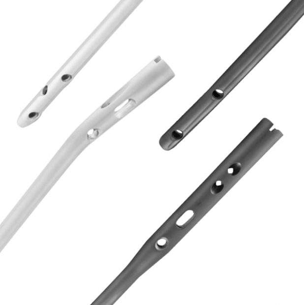
Treatment by balanced traction: Treatment by simple traction is suitable in many cases. The knee-joint can be made to exercise from the beginning. This is suitable for vertical, comminuted and Y-shaped fractures. Excessive depression of the fracture will require elevation by operation. Surgeons need various ortho surgical implants and surgical tools to perform the operation, which is provided by Orthopedic Implants Suppliers in Romania.
- Aspiration: The hemarthrosis is aspirated with aseptic measures.
- Compression: Manipulation can be done under general anesthesia when the sideward position is corrected by applying compression force with both hands.
- Traction: Balanced skeletal traction is applied on a Thomas splint with Pearson’s knee flexion piece attached.
- Post-immobilization period: The quadriceps and knee-joint exercises are performed from the beginning. The traction is removed after a period of 6 weeks when the union is usually complete.
- Ambulation: Non-weight bearing with crutches is allowed once the traction is removed. Weight bearing is permitted after 6 weeks.
Treatment in plaster: Treatment can also be done by applying a long leg plaster. The disadvantage of this technique is that early exercise of the knee-joint cannot be performed. After reduction, the long leg plaster is applied. This is usually removed after 6-12 weeks, depending upon the nature of fracture.
Operation: Operative reduction is required in case of failure of closed reduction, unstable fracture and cases of markedly depressed fracture.
Nature of operation: Operation is performed in the form of internal fixation by intramedullary nails or pin and plate. Bone graft is done to fill the gap during the process of elevation of depression fracture.c